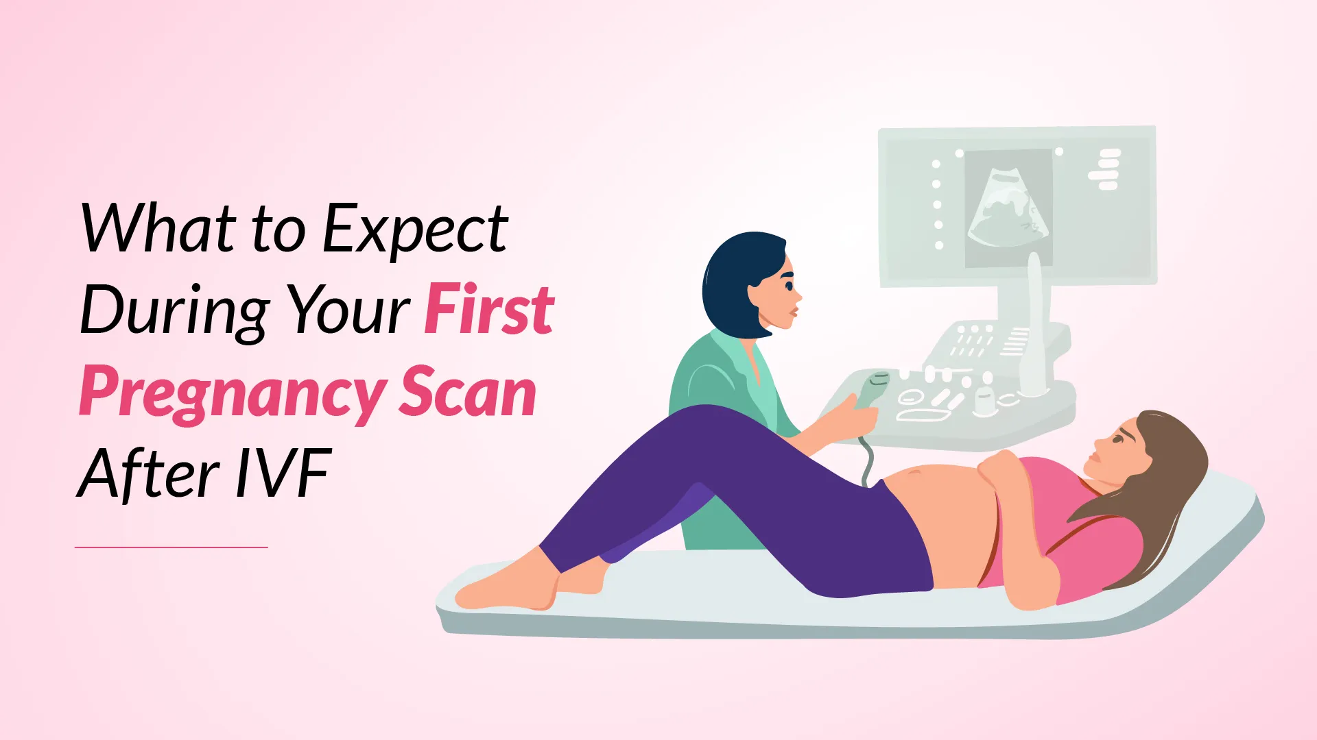
The first pregnancy scan after IVF marks a crucial milestone in the fertility journey. For many patients who have undergone IVF treatment, this scan represents the first visual confirmation of their pregnancy’s progress.
This guide explains everything patients need to know about their post-IVF pregnancy test, including when to schedule it, what happens during the appointment, and how to prepare both emotionally and practically for this significant moment.
Also read: How Soon Can a Pregnancy Test Be Taken After Implantation?
What Is the First Pregnancy Scan?
The 1st ultrasound after IVF is an early ultrasound examination that typically takes place between 6 to 8 weeks after embryo transfer. This initial scan, also known as a viability scan, is performed using a specialised transvaginal ultrasound device that provides detailed images of the developing pregnancy.
Doctors can gather essential information about the pregnancy’s progress during this crucial examination. The scan helps to:
- Confirm the presence of a gestational sac
- Verify the location of the pregnancy within the uterus
- Check for the presence of a foetal heartbeat
- Determine if there are multiple pregnancies
- Measure the size of the embryo
The first pregnancy scan differs from regular ultrasounds as it requires more precise monitoring due to the nature of IVF pregnancies. Doctors use high-frequency sound waves to create detailed images that can detect even the earliest signs of pregnancy development.
This early scan is essential for IVF patients as it provides the first visual confirmation of pregnancy success. Unlike standard pregnancy tests that only detect hormone levels, this ultrasound examination offers concrete evidence of embryo development and placement, helping to reassure patients about their pregnancy’s progression.
When to Schedule Your First Pregnancy Scan
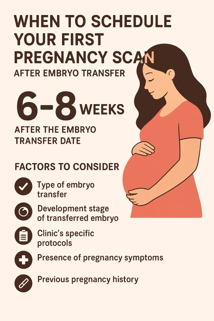
Scheduling the first pregnancy scan requires careful coordination with the fertility clinic. Most clinics recommend booking this necessary appointment approximately 6-8 weeks after the embryo transfer date.
Several factors determine the correct timing of the first scan:
- The type of embryo transfer performed (fresh or frozen)
- The development stage of the transferred embryo
- The clinic’s specific protocols
- The presence of any pregnancy symptoms
- Previous pregnancy history
Doctors typically schedule the scan at a point when they can expect to see clear signs of pregnancy development. This timing ensures that the ultrasound can provide meaningful information about the pregnancy’s progress. Patients should always follow their clinic’s specific guidelines, as protocols may vary between different fertility centres.
What Happens During the First Pregnancy Scan?
During the first pregnancy scan, patients will experience a thorough examination that typically takes about 20-30 minutes. The doctor will conduct the scan using a specialised transvaginal ultrasound probe, which provides the most explicit early pregnancy images.
The scan process involves several key components:
- The patient lies on an examination table in a comfortable position
- A specially trained sonographer performs the ultrasound
- The transvaginal probe is gently inserted to capture detailed images
- Real-time images appear on a monitor that both patient and doctor can view
- The sonographer takes specific measurements and captures important images
The Results of Your First Pregnancy Scan
Results from the first pregnancy scan provide crucial information that helps doctors assess the success of the IVF treatment. Understanding these outcomes helps patients prepare for the next steps in their fertility journey.
Positive Pregnancy Test After IVF
A positive scan result confirms the presence of a viable pregnancy. Doctors look for several key indicators:
- Clear visualisation of the gestational sac
- Presence of foetal pole
- Detection of cardiac activity
- Appropriate measurements for gestational age
- Correct placement in the uterus
With IVF positive results, the fertility team will schedule follow-up appointments and provide guidance for ongoing prenatal care. Patients typically transition to regular obstetric care around the 12-week mark.
For a more accurate prediction of your timeline, try our IVF Due Date Calculator to plan your next steps
Negative Pregnancy Test After IVF
When scan results don’t show the expected signs of pregnancy development, doctors will discuss the situation sensitively with patients. They will:
- Explain the findings in detail
- Discuss potential causes
- Outline available options for future treatment
- Provide emotional support resources
- Schedule follow-up consultations
Many fertility clinics have counsellors specialising in helping patients process their results, whether positive or negative.
Why Is the First Pregnancy Scan Important?
This vital examination serves multiple essential purposes that benefit both doctors and expectant parents.
The first scan plays a critical role in the following:
- Confirming the viability of the pregnancy
- Detecting potential complications early
- Providing precise dating of the pregnancy
- Assessing the need for additional medical support
- Offering visual confirmation of treatment success
- Helping doctors plan future care
Beyond its medical significance, the first pregnancy scan serves as a pivotal moment for emotional reassurance. Doctors can use this opportunity to address any concerns and adjust treatment plans if necessary. The detailed information gathered during this examination helps establish a solid foundation for ongoing prenatal care.
This scan holds particular importance for IVF patients as it confirms the successful implantation and development of the transferred embryo. The detailed images provide doctors with essential data about the pregnancy’s progression, allowing them to make informed decisions about subsequent care and monitoring requirements.
Emotional and Practical Preparation for the First Scan
Preparing for the first pregnancy scan after IVF involves both emotional and practical considerations. Many patients find themselves experiencing a mix of excitement and anxiety as they approach this significant milestone in their fertility journey.
Emotional preparation begins with acknowledging that feeling nervous is entirely normal. Doctors recommend that patients:
- Share their feelings with their partner or trusted friend
- Connect with IVF support groups or online communities
- Practise relaxation techniques before the appointment
- Prepare questions for the healthcare team
- Consider bringing a support person to the scan
- Set realistic expectations about the appointment
For in-clinic preparation, patients should ensure they have all necessary documentation and follow any specific instructions from their fertility clinic. It’s helpful to wear comfortable clothing and reach the clinic with a comfortably full bladder, as this can help with image clarity during the scan.
Many fertility clinics provide dedicated support services, including counselling, to help patients manage their emotions during this time.
Find Hope and Solutions for Female Infertility and Male Infertility — Explore Our Comprehensive Services
IUI Treatment
ICSI Treatment
PICSI Treatment
Fertility Preservation Service
Blastocyst Culture & Transfer Treatment
Genetic Screening & Testing
Conclusion
The first pregnancy scan after IVF marks a significant milestone that brings both medical insights and emotional reassurance. This crucial examination helps doctors confirm pregnancy viability while giving expectant parents their first glimpse of their developing baby.
Patients should remember that feeling nervous before the scan is natural and normal. The healthcare team understands these concerns and provides support throughout the ivf process steps. Most fertility clinics offer comprehensive guidance, from scheduling the right time for the scan to helping patients process the results.
The success of IVF treatment becomes more evident through this initial scan, which typically happens between 6 to 8 weeks after embryo transfer. Doctors use this detailed examination to check the embryo’s placement, development, and heartbeat, setting the foundation for future prenatal care.
A well-prepared approach to the first pregnancy scan, both emotionally and practically, helps patients make the most of this important appointment. The support of doctors and a clear understanding of what to expect can make this milestone moment less stressful and more meaningful for everyone involved.
